 국립중앙도서관 "우편 복사 서비스"로 연결 됩니다.


 KCI
KCIProbe-based confocal microscopy (pCLE) is actively being investigated for applications in the esophagus and stomach. The use of pCLE allows real-time in vivo microscopy to evaluate the microarchitecture of the mucosal epithelium. pCLE appears to be pa...
다국어 입력
あ
ぁ
か
が
さ
ざ
た
だ
な
は
ば
ぱ
ま
や
ゃ
ら
わ
ゎ
ん
い
ぃ
き
ぎ
し
じ
ち
ぢ
に
ひ
び
ぴ
み
り
う
ぅ
く
ぐ
す
ず
つ
づ
っ
ぬ
ふ
ぶ
ぷ
む
ゆ
ゅ
る
え
ぇ
け
げ
せ
ぜ
て
で
ね
へ
べ
ぺ
め
れ
お
ぉ
こ
ご
そ
ぞ
と
ど
の
ほ
ぼ
ぽ
も
よ
ょ
ろ
を
ア
ァ
カ
サ
ザ
タ
ダ
ナ
ハ
バ
パ
マ
ヤ
ャ
ラ
ワ
ヮ
ン
イ
ィ
キ
ギ
シ
ジ
チ
ヂ
ニ
ヒ
ビ
ピ
ミ
リ
ウ
ゥ
ク
グ
ス
ズ
ツ
ヅ
ッ
ヌ
フ
ブ
プ
ム
ユ
ュ
ル
エ
ェ
ケ
ゲ
セ
ゼ
テ
デ
ヘ
ベ
ペ
メ
レ
オ
ォ
コ
ゴ
ソ
ゾ
ト
ド
ノ
ホ
ボ
ポ
モ
ヨ
ョ
ロ
ヲ
―
http://chineseinput.net/에서 pinyin(병음)방식으로 중국어를 변환할 수 있습니다.
변환된 중국어를 복사하여 사용하시면 됩니다.
예시)
- 中文 을 입력하시려면 zhongwen을 입력하시고 space를누르시면됩니다.
- 北京 을 입력하시려면 beijing을 입력하시고 space를 누르시면 됩니다.
А
Б
В
Г
Д
Е
Ё
Ж
З
И
Й
К
Л
М
Н
О
П
Р
С
Т
У
Ф
Х
Ц
Ч
Ш
Щ
Ъ
Ы
Ь
Э
Ю
Я
а
б
в
г
д
е
ё
ж
з
и
й
к
л
м
н
о
п
р
с
т
у
ф
х
ц
ч
ш
щ
ъ
ы
ь
э
ю
я
′
″
℃
Å
¢
£
¥
¤
℉
‰
$
%
F
₩
㎕
㎖
㎗
ℓ
㎘
㏄
㎣
㎤
㎥
㎦
㎙
㎚
㎛
㎜
㎝
㎞
㎟
㎠
㎡
㎢
㏊
㎍
㎎
㎏
㏏
㎈
㎉
㏈
㎧
㎨
㎰
㎱
㎲
㎳
㎴
㎵
㎶
㎷
㎸
㎹
㎀
㎁
㎂
㎃
㎄
㎺
㎻
㎽
㎾
㎿
㎐
㎑
㎒
㎓
㎔
Ω
㏀
㏁
㎊
㎋
㎌
㏖
㏅
㎭
㎮
㎯
㏛
㎩
㎪
㎫
㎬
㏝
㏐
㏓
㏃
㏉
㏜
㏆

RISS 인기검색어
https://www.riss.kr/link?id=A101604825
-
저자
Adam Templeton (Division of Gastroenterology,Department of Medicine,University of Washington,Seattle,USA) ; 황주하 (워싱턴대학교)
- 발행기관
- 학술지명
- 권호사항
-
발행연도
2013
-
작성언어
English
- 주제어
-
등재정보
KCI등재,SCOPUS,ESCI
-
자료형태
학술저널
- 발행기관 URL
-
수록면
445-449(5쪽)
-
KCI 피인용횟수
3
- 제공처
-
0
상세조회 -
0
다운로드
부가정보
다국어 초록 (Multilingual Abstract)
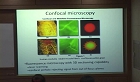
Probe-based confocal microscopy (pCLE) is actively being investigated for applications in the esophagus and stomach. The use of pCLE allows real-time in vivo microscopy to evaluate the microarchitecture of the mucosal epithelium. pCLE appears to be particularly useful in identifying mucosal dysplasia and early malignancies that cannot be clearly distinguished using high-definition white light endoscopy, chromoendoscopy, or magnification endoscopy. In addition, the ability to detect dysplastic tissue in real-time may shift the current screening practice from random biopsy to targeted biopsy of esophageal and gastric cancers and their precursor lesions. We will review the use of pCLE for detection and surveillance of upper gastrointestinal early luminal malignancy.
참고문헌 (Reference)
1 Wallace MB, "The safety of intravenous fluorescein for confocal laser endomicroscopy in the gastrointestinal tract" 31 : 548-552, 2010
2 ASGE Standards of Practice Committee, "The role of endoscopy in Barrett’s esophagus and other premalignant conditions of the esophagus" 76 : 1087-1094, 2012
3 Pittayanon R, "The learning curve of gastric intestinal metaplasia interpretation on the images obtained by probe-based confocal laser endomicroscopy" 2012 : 278045-, 2012
4 Bok GH, "The accuracy of probe-based confocal endomicroscopy versus conventional endoscopic biopsies for the diagnosis of superficial gastric neoplasia (with videos)" 77 : 899-908, 2013
5 Gupta M, "Recurrence of esophageal intestinal metaplasia after endoscopic mucosal resection and radiofrequency ablation of Barrett’s esophagus: results from a US Multicenter Consortium" 145 : 79-86, 2013
6 Sharma P, "Real-time increased detection of neoplastic tissue in Barrett’s esophagus with probe-based confocal laser endomicroscopy: final results of an international multicenter, prospective, randomized, controlled trial" 74 : 465-472, 2011
7 Bajbouj M, "Probe-based confocal laser endomicroscopy compared with standard four-quadrant biopsy for evaluation of neoplasia in Barrett’s esophagus" 42 : 435-440, 2010
8 Wallace MB, "Probe-based confocal laser endomicroscopy" 136 : 1509-1513, 2009
9 Gaddam S, "Novel probe-based confocal laser endomicroscopy criteria and interobserver agreement for the detection of dysplasia in Barrett’s esophagus" 106 : 1961-1969, 2011
10 Capelle LG, "Narrow band imaging for the detection of gastric intestinal metaplasia and dysplasia during surveillance endoscopy" 55 : 3442-3448, 2010
11 Wallace MB, "Multicenter, randomized, controlled trial of confocal laser endomicroscopy assessment of residual metaplasia after mucosal ablation or resection of GI neoplasia in Barrett’s esophagus" 76 : 539-547, 2012
12 Wallace M, "Miami classification for probebased confocal laser endomicroscopy" 43 : 882-891, 2011
13 Kato M, "Magnifying endoscopy with narrow- band imaging achieves superior accuracy in the differential diagnosis of superficial gastric lesions identified with white-light endoscopy: a prospective study" 72 : 523-529, 2010
14 Uedo N, "Longterm outcomes after endoscopic mucosal resection for early gastric cancer" 9 : 88-92, 2006
15 Wang TD, "Functional imaging of colonic mucosa with a fibered confocal microscope for real-time in vivo pathology" 5 : 1300-1305, 2007
16 Shaheen NJ, "Durability of radiofrequency ablation in Barrett’s esophagus with dysplasia" 141 : 460-468, 2011
17 Gorospe EC, "Diagnostic performance of two confocal endomicroscopy systems in detecting Barrett’s dysplasia: a pilot study using a novel bioprobe in ex vivo tissue" 76 : 933-938, 2012
18 Guo YT, "Diagnosis of gastric intestinal metaplasia with confocal laser endomicroscopy in vivo: a prospective study" 40 : 547-553, 2008
19 Kiesslich R, "Confocal laser endoscopy for diagnosing intraepithelial neoplasias and colorectal cancer in vivo" 127 : 706-713, 2004
20 American Gastroenterological Association, "American Gastroenterological Association medical position statement on the management of Barrett’s esophagus" 140 : 1084-1091, 2011
21 Abrams JA, "Adherence to biopsy guidelines for Barrett’s esophagus surveillance in the community setting in the United States" 7 : 736-742, 2009
22 Polglase AL, "A fluorescence confocal endomicroscope for in vivo microscopy of the upper- and the lower-GI tract" 62 : 686-695, 2005
동일학술지(권/호) 다른 논문
-
Systemic Amyloidosis Manifested by Gastric Outlet Obstruction
- 대한소화기내시경학회
- 박성운
- 2013
- KCI등재,SCOPUS,ESCI
-
Successful Endoscopic Mucosal Resection of a Low Esophageal Carcinoid Tumor
- 대한소화기내시경학회
- 임창섭
- 2013
- KCI등재,SCOPUS,ESCI
-
Gastrointestinal Cancers in a Peutz–Jeghers Syndrome family: A Case Report
- 대한소화기내시경학회
- 송상희
- 2013
- KCI등재,SCOPUS,ESCI
-
- 대한소화기내시경학회
- 이상진
- 2013
- KCI등재,SCOPUS,ESCI
분석정보
인용정보 인용지수 설명보기
학술지 이력
| 연월일 | 이력구분 | 이력상세 | 등재구분 |
|---|---|---|---|
| 2023 | 평가예정 | 해외DB학술지평가 신청대상 (해외등재 학술지 평가) | |
| 2020-01-01 | 평가 | 등재학술지 유지 (해외등재 학술지 평가) |  |
| 2011-12-21 | 학술지명변경 | 한글명 : 대한소화기내시경학회지 -> Clinical Endoscopy외국어명 : The Korean Journal of Gastrointestinal Endoscopy -> Clinical Endoscopy | |
| 2010-01-01 | 평가 | 등재학술지 유지 (등재유지) | |
| 2007-01-01 | 평가 | 등재학술지 선정 (등재후보2차) | |
| 2006-06-22 | 학술지명변경 | 한글명 : 대한소화기내시경학회 -> 대한소화기내시경학회지 |  |
| 2006-06-21 | 학술지등록 | 한글명 : 대한소화기내시경학회외국어명 : The Korean Journal of Gastrointestinal Endoscopy | |
| 2006-01-01 | 평가 | 등재후보 1차 PASS (등재후보1차) | |
| 2004-07-01 | 평가 | 등재후보학술지 선정 (신규평가) | |
학술지 인용정보
| 기준연도 | WOS-KCI 통합IF(2년) | KCIF(2년) | KCIF(3년) |
|---|---|---|---|
| 2016 | 0.23 | 0.22 | 0.23 |
| KCIF(4년) | KCIF(5년) | 중심성지수(3년) | 즉시성지수 |
| 0.21 | 0.18 | 0.38 | 0.25 |
이 자료와 함께 이용한 RISS 자료
나만을 위한 추천자료


