 국립중앙도서관 "우편 복사 서비스"로 연결 됩니다.

 KCI
KCIStudy Design: This is an in-vitro experimental study Objectives: We wanted to analyze the changes in the growth and phenotype of human degenerative intervertebral disc cells depending on the frequency of subculture in an in vitro monolayer culture sys...
다국어 입력
あ
ぁ
か
が
さ
ざ
た
だ
な
は
ば
ぱ
ま
や
ゃ
ら
わ
ゎ
ん
い
ぃ
き
ぎ
し
じ
ち
ぢ
に
ひ
び
ぴ
み
り
う
ぅ
く
ぐ
す
ず
つ
づ
っ
ぬ
ふ
ぶ
ぷ
む
ゆ
ゅ
る
え
ぇ
け
げ
せ
ぜ
て
で
ね
へ
べ
ぺ
め
れ
お
ぉ
こ
ご
そ
ぞ
と
ど
の
ほ
ぼ
ぽ
も
よ
ょ
ろ
を
ア
ァ
カ
サ
ザ
タ
ダ
ナ
ハ
バ
パ
マ
ヤ
ャ
ラ
ワ
ヮ
ン
イ
ィ
キ
ギ
シ
ジ
チ
ヂ
ニ
ヒ
ビ
ピ
ミ
リ
ウ
ゥ
ク
グ
ス
ズ
ツ
ヅ
ッ
ヌ
フ
ブ
プ
ム
ユ
ュ
ル
エ
ェ
ケ
ゲ
セ
ゼ
テ
デ
ヘ
ベ
ペ
メ
レ
オ
ォ
コ
ゴ
ソ
ゾ
ト
ド
ノ
ホ
ボ
ポ
モ
ヨ
ョ
ロ
ヲ
―
http://chineseinput.net/에서 pinyin(병음)방식으로 중국어를 변환할 수 있습니다.
변환된 중국어를 복사하여 사용하시면 됩니다.
예시)
- 中文 을 입력하시려면 zhongwen을 입력하시고 space를누르시면됩니다.
- 北京 을 입력하시려면 beijing을 입력하시고 space를 누르시면 됩니다.
А
Б
В
Г
Д
Е
Ё
Ж
З
И
Й
К
Л
М
Н
О
П
Р
С
Т
У
Ф
Х
Ц
Ч
Ш
Щ
Ъ
Ы
Ь
Э
Ю
Я
а
б
в
г
д
е
ё
ж
з
и
й
к
л
м
н
о
п
р
с
т
у
ф
х
ц
ч
ш
щ
ъ
ы
ь
э
ю
я
′
″
℃
Å
¢
£
¥
¤
℉
‰
$
%
F
₩
㎕
㎖
㎗
ℓ
㎘
㏄
㎣
㎤
㎥
㎦
㎙
㎚
㎛
㎜
㎝
㎞
㎟
㎠
㎡
㎢
㏊
㎍
㎎
㎏
㏏
㎈
㎉
㏈
㎧
㎨
㎰
㎱
㎲
㎳
㎴
㎵
㎶
㎷
㎸
㎹
㎀
㎁
㎂
㎃
㎄
㎺
㎻
㎽
㎾
㎿
㎐
㎑
㎒
㎓
㎔
Ω
㏀
㏁
㎊
㎋
㎌
㏖
㏅
㎭
㎮
㎯
㏛
㎩
㎪
㎫
㎬
㏝
㏐
㏓
㏃
㏉
㏜
㏆

RISS 인기검색어

세포 치료를 위한 인간 추간판 세포의 적절한 체외 계대 배양 횟수 -세포의 성장 및 표현형- = Adequate Serial Monolayer Passage Number of Human Intervertebral Disc Cells for Cell Therapy -Growth and Phenotype of Cells-
한글로보기https://www.riss.kr/link?id=A104778317
- 저자
- 발행기관
- 학술지명
- 권호사항
-
발행연도
2010
-
작성언어
Korean
-
주제어
추간판 ; 퇴행성 변화 ; 표현형 ; 계대 배양 ; Intervertebral disc ; Degenerative change ; Phenotype ; Subculture
-
등재정보
KCI등재
-
자료형태
학술저널
- 발행기관 URL
-
수록면
57-65(9쪽)
-
KCI 피인용횟수
0
- 제공처
-
0
상세조회 -
0
다운로드
부가정보
다국어 초록 (Multilingual Abstract)

Study Design: This is an in-vitro experimental study Objectives: We wanted to analyze the changes in the growth and phenotype of human degenerative intervertebral disc cells depending on the frequency of subculture in an in vitro monolayer culture system.
Summary of the Literature Review: A subculture of disc cells is needed to obtain an adequate amount of disc cells for cell therapy,tissue engineering and analysis of the biological characteristics of degenerative disc cells Materials and Methods: The obtained intervertebral discs were divided into the nucleus pulposus (NP) and the annulus fibrosus (AF).
The AF and NP cells were cultured in a monolayer manner, respectively. At each subculture time, we analyzed the morphological changes,the adhesion rate, the proliferation rate and the viability. The expressions of types I and II collagen and proteoglycan were analyzed at the mRNA gene level.
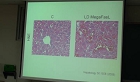
Results: Both the AF and NP cells gradually showed a fibroblast-like spindle shape while undergoing subculture. The adhesion rate was higher at the second and third times of subculture. The cell proliferation was the highest at the second subculture time. The viability was markedly lower prior to the subculture. On RT-PCR, the type II collagen expression was gradually decreased in the NP cells. In the AF cells, Type II collagen was not expressed from the second time of subculture. The expression of proteoglycan was gradually decreased in both.
Conclusions: Following the 3rd subculture, the degenerative disc cells had completely changed their original growth and phenotypic characteristics. Therefore, we believe that it is not desirable for us to do passage cultures more than three times for cell therapy.
Summary of the Literature Review: A subculture of disc cells is needed to obtain an adequate amount of disc cells for cell therapy,tissue engineering and analysis of the biological characteristics of degenerative disc cells Materials and Methods: The obtained intervertebral discs were divided into the nucleus pulposus (NP) and the annulus fibrosus (AF).
The AF and NP cells were cultured in a monolayer manner, respectively. At each subculture time, we analyzed the morphological changes,the adhesion rate, the proliferation rate and the viability. The expressions of types I and II collagen and proteoglycan were analyzed at the mRNA gene level.
Results: Both the AF and NP cells gradually showed a fibroblast-like spindle shape while undergoing subculture. The adhesion rate was higher at the second and third times of subculture. The cell proliferation was the highest at the second subculture time. The viability was markedly lower prior to the subculture. On RT-PCR, the type II collagen expression was gradually decreased in the NP cells. In the AF cells, Type II collagen was not expressed from the second time of subculture. The expression of proteoglycan was gradually decreased in both.
Conclusions: Following the 3rd subculture, the degenerative disc cells had completely changed their original growth and phenotypic characteristics. Therefore, we believe that it is not desirable for us to do passage cultures more than three times for cell therapy.
Study Design: This is an in-vitro experimental study Objectives: We wanted to analyze the changes in the growth and phenotype of human degenerative intervertebral disc cells depending on the frequency of subculture in an in vitro monolayer culture system.
Summary of the Literature Review: A subculture of disc cells is needed to obtain an adequate amount of disc cells for cell therapy,tissue engineering and analysis of the biological characteristics of degenerative disc cells Materials and Methods: The obtained intervertebral discs were divided into the nucleus pulposus (NP) and the annulus fibrosus (AF).
The AF and NP cells were cultured in a monolayer manner, respectively. At each subculture time, we analyzed the morphological changes,the adhesion rate, the proliferation rate and the viability. The expressions of types I and II collagen and proteoglycan were analyzed at the mRNA gene level.
Results: Both the AF and NP cells gradually showed a fibroblast-like spindle shape while undergoing subculture. The adhesion rate was higher at the second and third times of subculture. The cell proliferation was the highest at the second subculture time. The viability was markedly lower prior to the subculture. On RT-PCR, the type II collagen expression was gradually decreased in the NP cells. In the AF cells, Type II collagen was not expressed from the second time of subculture. The expression of proteoglycan was gradually decreased in both.
Conclusions: Following the 3rd subculture, the degenerative disc cells had completely changed their original growth and phenotypic characteristics. Therefore, we believe that it is not desirable for us to do passage cultures more than three times for cell therapy.
국문 초록 (Abstract)
연구 계획: 시험관 실험적 연구목적: 사람의 퇴행성 추간판 세포를 체외에서 배양하면서 계대 배양의 횟수에 따른 성장 및 표현형의 변화를 분석하고자 한다.
선행문헌의 요약: 세포 치료, 조직공학 및 추간판 세포의 생물학적 특성의 분석을 위해서는 퇴행성 추간판 세포의 계대 배양을 거쳐 많은 양의 세포를 획득하여야 한다.
대상 및 방법:. 적출된 추간판 조직을 수핵과 섬유륜 부분으로 나누어서 단층 배양하였다. 계대 배양을 거치면서 세포의 형태 변화, 부착율, 증식율 및 생존율 분석을 진행하였다. 또한 RT-PCR 검사를 시행하여 제 1, 2형 교원질 (type I, II collagen) 및 단백다당 (proteoglycan) mRNA 발현을 유전자 수준에서 분석하였다.
결과: 세포의 모양은 수핵 및 섬유륜 세포 모두 점차적으로 방추형인 섬유모세포 모양을 나타냈으며, 부착율은 2차 및 3차 계대 배양에서 상대적으로 증가하였고, 증식율은 2차 계대 배양에서 가장 우수했으며, 세포의 생존율은 배양을 거치기 전에는 현저히 낮았다. RT-PCR 분석 상 수핵 세포의 제2형 교원질 발현은 점차적으로 감소하는 양상을 보였으며, 섬유륜 세포의 제 2형 교원질은 2차 계대 배양 시부터 발현이 되지 않았다. 단백 다당의 발현은 수핵 및 섬유륜 세포 모두에서 점차 감소하는 양상을 보였다.
결론: 퇴행성 변화를 일으킨 추간판의 섬유륜 및 수핵 세포는 체외에서 계대 배양의 횟수가 3차 계대 배양에서는 고유의 성장과 표현형을 잃어버렸다. 이에 세포치료에 있어서 체외에서 단층 배양 시 3번 이상의 계대 배양을 거치지 않는 것이 바람직할 것으로 사료된다.
연구 계획: 시험관 실험적 연구목적: 사람의 퇴행성 추간판 세포를 체외에서 배양하면서 계대 배양의 횟수에 따른 성장 및 표현형의 변화를 분석하고자 한다. 선행문헌의 요약: 세포 치료, ...
연구 계획: 시험관 실험적 연구목적: 사람의 퇴행성 추간판 세포를 체외에서 배양하면서 계대 배양의 횟수에 따른 성장 및 표현형의 변화를 분석하고자 한다.
선행문헌의 요약: 세포 치료, 조직공학 및 추간판 세포의 생물학적 특성의 분석을 위해서는 퇴행성 추간판 세포의 계대 배양을 거쳐 많은 양의 세포를 획득하여야 한다.
대상 및 방법:. 적출된 추간판 조직을 수핵과 섬유륜 부분으로 나누어서 단층 배양하였다. 계대 배양을 거치면서 세포의 형태 변화, 부착율, 증식율 및 생존율 분석을 진행하였다. 또한 RT-PCR 검사를 시행하여 제 1, 2형 교원질 (type I, II collagen) 및 단백다당 (proteoglycan) mRNA 발현을 유전자 수준에서 분석하였다.
결과: 세포의 모양은 수핵 및 섬유륜 세포 모두 점차적으로 방추형인 섬유모세포 모양을 나타냈으며, 부착율은 2차 및 3차 계대 배양에서 상대적으로 증가하였고, 증식율은 2차 계대 배양에서 가장 우수했으며, 세포의 생존율은 배양을 거치기 전에는 현저히 낮았다. RT-PCR 분석 상 수핵 세포의 제2형 교원질 발현은 점차적으로 감소하는 양상을 보였으며, 섬유륜 세포의 제 2형 교원질은 2차 계대 배양 시부터 발현이 되지 않았다. 단백 다당의 발현은 수핵 및 섬유륜 세포 모두에서 점차 감소하는 양상을 보였다.
결론: 퇴행성 변화를 일으킨 추간판의 섬유륜 및 수핵 세포는 체외에서 계대 배양의 횟수가 3차 계대 배양에서는 고유의 성장과 표현형을 잃어버렸다. 이에 세포치료에 있어서 체외에서 단층 배양 시 3번 이상의 계대 배양을 거치지 않는 것이 바람직할 것으로 사료된다.
참고문헌 (Reference)
1 Bayliss MT, "Volvo award in basic science. Proteoglycan synthesis in the human intervertebral disc. Variation with age, region and pathology" 13 : 972-981, 1988
2 Brittberg M, "Treatment of deep cartilage defects in the knee with autologous chondrocyte transplantation" 331 : 889-895, 1994
3 Freeman BJ, "Total disc replacement in the lumbar spine: a systematic review of the literature" 15 : 439-447, 2006
4 Johnstone B, "The large proteoglycans of the human intervertebral disc. Changes in their biosynthesis and structure with age, topography, and pathology" 20 : 674-684, 1995
5 Eie N, "The knee-elbow position in lumbar disc surgery: a review of complications" 8 : 897-900, 1983
6 Chou AI, "The effect of serial monolayer passaging on the collagen expression profile of outer and inner anulus fibrosus cells" 31 : 1875-1881, 2006
7 Eyre DR, "Quantitative analysis of types I and II collagens in human intervertebral discs at various ages" 492 : 29-42, 1977
8 Yeung AT, "Posterolateral endoscopic excision for lumbar disc herniation: Surgical technique, outcome, and complications in 307 consecutive cases" 27 : 722-731, 2002
9 Hoppenfeld S, "Percutaneous removal of herniated lumbar discs. 50 cases with ten-year follow-up periods" 238 : 92-97, 1989
10 Lee JY, "New use of a three-dimensional pellet culture system for human intervertebral disc cells: initial characterization and potential use for tissue engineering" 26 : 2316-2322, 2001
11 Yoon ST, "Molecular therapy of the intervertebral disc" 5 : 280-286, 2005
12 White AH, "Lumbar laminectomy for herniated disc: a prospective controlled comparison with internal fixation fusion" 12 : 305-307, 1987
13 Gan JC, "Intervertebral disc tissue engineering I: characterization of the nucleus pulposus" 411 : 305-314, 2003
14 Wang JY, "Intervertebral disc cells exhibit differences in gene expression in alginate and monolayer culture" 26 : 1747-1751, 2001
15 Loeser RF, "Integrin expression by primary and immortalized human chondrocytes: evidence of a differential role for alpha1beta1 and alpha2beta1 integrins in mediating chondrocyte adhesion to types II and VI collagen" 8 : 96-105, 2000
16 Tullberg-Reinert H, "In situ measurement of collagen synthesis by human bone cells with a sirius red-based colorimetric microassay: effects of transforming growth factor beta2 and ascorbic acid 2-phosphate" 112 : 271-276, 1999
17 Farndale RW, "Improved quantitation and discrimination of sulphated glycosaminoglycans by use of dimethylmethylene blue" 883 : 173-177, 1986
18 Kluba T, "Human anulus fibrosus and nucleus pulposus cells of the intervertebral disc: effect of degeneration and culture system on cell phenotype" 30 : 2743-2748, 2005
19 Yashiki S, "Evaluation of attachment and growth of anchorage-dependent cells on culture surfaces with type I collagen coating" 92 : 385-388, 2001
20 Urban JP, "Degeneration of the intervertebral disc" 5 : 120-130, 2003
21 Benya PD, "Dedifferentiated chondrocytes reexpress the differentiated collagen phenotype when cultured in agarose gels" 30 : 215-224, 1982
22 Lemare F, "Dedifferentiated chondrocytes cultured in alginate beads: restoration of the differentiated phenotype and of the metabolic responses to interleukin-1beta" 176 : 303-313, 1998
23 Freemont AJ, "Current understanding of cellular and molecular events in intervertebral disc degeneration: implications for therapy" 196 : 374-379, 2002
24 Eie N, "Comparison of the results in patients operated upon for ruptured lumbar discs with and without spinal fusion" 41 : 107-113, 1978
25 Hong Y, "Collagen-coated polylactide microspheres as chondrocyte microcarriers" 26 : 6305-6313, 2005
26 Wang H, "Chondrocytes attach to hyaline or calcified cartilage and bone" 12 : 56-64, 2004
27 An HS, "Biological repair of intervertebral disc" 28 : 86-92, 2003
28 Baums MH, "Autologous chondrocyte transplantation for treating cartilage defects of the talus" 88 : 303-308, 2006
29 Sato M, "An atelocollagen honeycomb-shaped scaffold with a membrane seal (ACHMS-scaffold) for the culture of annulus fibrosus cells from an intervertebral disc" 64 : 248-256, 2003
30 Ganey TM, "A potential role for cell-based therapeutics in the treatment of intervertebral disc herniation" 11 : 206-214, 2002
동일학술지(권/호) 다른 논문
-
투석을 시행하는 퇴행성 요추 질환 환자군에서의 수 술적 치료의 임상결과
- 대한척추외과학회
- 이병호
- 2010
- KCI등재
-
요추부 인접 분절 질환에 대한 위험 인자 분석 및 수 술 후 결과
- 대한척추외과학회
- 김환정
- 2010
- KCI등재
-
아텔로콜라겐 지지체, 유전자 치료, 배양된 수핵세포를 이용한 조직 공학적 추간판의 재생
- 대한척추외과학회
- 김학선
- 2010
- KCI등재
-
정상 한국인의 요추 형태 분류에 따른 시상면상 척 추 - 골반 지표들의 변화 -20대 성인 남자 군에서 의 연구-
- 대한척추외과학회
- 안영준
- 2010
- KCI등재
분석정보
인용정보 인용지수 설명보기
학술지 이력
| 연월일 | 이력구분 | 이력상세 | 등재구분 |
|---|---|---|---|
| 2027 | 평가예정 | 재인증평가 신청대상 (재인증) | |
| 2021-01-01 | 평가 | 등재학술지 유지 (재인증) | |
| 2018-01-01 | 평가 | 등재학술지 선정 (계속평가) | |
| 2017-12-01 | 평가 | 등재후보로 하락 (계속평가) |  |
| 2013-01-01 | 평가 | 등재 1차 FAIL (등재유지) | |
| 2010-01-01 | 평가 | 등재학술지 선정 (등재후보2차) | |
| 2009-01-01 | 평가 | 등재후보 1차 PASS (등재후보1차) | |
| 2007-01-01 | 평가 | 등재후보학술지 선정 (신규평가) | |
학술지 인용정보
| 기준연도 | WOS-KCI 통합IF(2년) | KCIF(2년) | KCIF(3년) |
|---|---|---|---|
| 2016 | 0.03 | 0.03 | 0.04 |
| KCIF(4년) | KCIF(5년) | 중심성지수(3년) | 즉시성지수 |
| 0.06 | 0.05 | 0.228 | 0 |


이 자료와 함께 이용한 RISS 자료
나만을 위한 추천자료


