 국립중앙도서관 "우편 복사 서비스"로 연결 됩니다.

 스콜라
스콜라Five dogs were submitted for diagnosis in Pathology Division of the National Veterinary Research Institute. These dogs showed emaciation and rough hair coat. At necropsy, numerous sharply demarcated yellowish white spots or streaks were scattered in ...
다국어 입력
あ
ぁ
か
が
さ
ざ
た
だ
な
は
ば
ぱ
ま
や
ゃ
ら
わ
ゎ
ん
い
ぃ
き
ぎ
し
じ
ち
ぢ
に
ひ
び
ぴ
み
り
う
ぅ
く
ぐ
す
ず
つ
づ
っ
ぬ
ふ
ぶ
ぷ
む
ゆ
ゅ
る
え
ぇ
け
げ
せ
ぜ
て
で
ね
へ
べ
ぺ
め
れ
お
ぉ
こ
ご
そ
ぞ
と
ど
の
ほ
ぼ
ぽ
も
よ
ょ
ろ
を
ア
ァ
カ
サ
ザ
タ
ダ
ナ
ハ
バ
パ
マ
ヤ
ャ
ラ
ワ
ヮ
ン
イ
ィ
キ
ギ
シ
ジ
チ
ヂ
ニ
ヒ
ビ
ピ
ミ
リ
ウ
ゥ
ク
グ
ス
ズ
ツ
ヅ
ッ
ヌ
フ
ブ
プ
ム
ユ
ュ
ル
エ
ェ
ケ
ゲ
セ
ゼ
テ
デ
ヘ
ベ
ペ
メ
レ
オ
ォ
コ
ゴ
ソ
ゾ
ト
ド
ノ
ホ
ボ
ポ
モ
ヨ
ョ
ロ
ヲ
―
http://chineseinput.net/에서 pinyin(병음)방식으로 중국어를 변환할 수 있습니다.
변환된 중국어를 복사하여 사용하시면 됩니다.
예시)
- 中文 을 입력하시려면 zhongwen을 입력하시고 space를누르시면됩니다.
- 北京 을 입력하시려면 beijing을 입력하시고 space를 누르시면 됩니다.
А
Б
В
Г
Д
Е
Ё
Ж
З
И
Й
К
Л
М
Н
О
П
Р
С
Т
У
Ф
Х
Ц
Ч
Ш
Щ
Ъ
Ы
Ь
Э
Ю
Я
а
б
в
г
д
е
ё
ж
з
и
й
к
л
м
н
о
п
р
с
т
у
ф
х
ц
ч
ш
щ
ъ
ы
ь
э
ю
я
′
″
℃
Å
¢
£
¥
¤
℉
‰
$
%
F
₩
㎕
㎖
㎗
ℓ
㎘
㏄
㎣
㎤
㎥
㎦
㎙
㎚
㎛
㎜
㎝
㎞
㎟
㎠
㎡
㎢
㏊
㎍
㎎
㎏
㏏
㎈
㎉
㏈
㎧
㎨
㎰
㎱
㎲
㎳
㎴
㎵
㎶
㎷
㎸
㎹
㎀
㎁
㎂
㎃
㎄
㎺
㎻
㎽
㎾
㎿
㎐
㎑
㎒
㎓
㎔
Ω
㏀
㏁
㎊
㎋
㎌
㏖
㏅
㎭
㎮
㎯
㏛
㎩
㎪
㎫
㎬
㏝
㏐
㏓
㏃
㏉
㏜
㏆

RISS 인기검색어

개 capillaria hepatica 感染 例의 病理學的 觀察 = Pathological findings of capillaria hepatica infection in dogs
한글로보기부가정보
다국어 초록 (Multilingual Abstract)
Five dogs were submitted for diagnosis in Pathology Division of the National Veterinary Research Institute. These dogs showed emaciation and rough hair coat.
At necropsy, numerous sharply demarcated yellowish white spots or streaks were scattered in the liver parenchyma under the capsule.
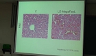
Histopathological findings were seen with typical granuloma in the liver parenchyma. Nematodes and their ova were surrounded by macrophages, epithelioid cells, foreign body giant cells, eosinophiles and lymphocytes, and these granulomatous nodules were encapsulated with fibrous connective tissues. The ova were lemon shaped, bipolar operculate and enveloped by the thick bilayered walls. The transparent outer wall had a radially striated appearance, whereas the inner wall was more dense and opaque. The mean dimension of ova were 53.5㎛ long by 28㎛ wide.
These results were concluded that five dogs were infected with Capillaria hepatica.
At necropsy, numerous sharply demarcated yellowish white spots or streaks were scattered in the liver parenchyma under the capsule.
Histopathological findings were seen with typical granuloma in the liver parenchyma. Nematodes and their ova were surrounded by macrophages, epithelioid cells, foreign body giant cells, eosinophiles and lymphocytes, and these granulomatous nodules were encapsulated with fibrous connective tissues. The ova were lemon shaped, bipolar operculate and enveloped by the thick bilayered walls. The transparent outer wall had a radially striated appearance, whereas the inner wall was more dense and opaque. The mean dimension of ova were 53.5㎛ long by 28㎛ wide.
These results were concluded that five dogs were infected with Capillaria hepatica.
Five dogs were submitted for diagnosis in Pathology Division of the National Veterinary Research Institute. These dogs showed emaciation and rough hair coat.
At necropsy, numerous sharply demarcated yellowish white spots or streaks were scattered in the liver parenchyma under the capsule.
Histopathological findings were seen with typical granuloma in the liver parenchyma. Nematodes and their ova were surrounded by macrophages, epithelioid cells, foreign body giant cells, eosinophiles and lymphocytes, and these granulomatous nodules were encapsulated with fibrous connective tissues. The ova were lemon shaped, bipolar operculate and enveloped by the thick bilayered walls. The transparent outer wall had a radially striated appearance, whereas the inner wall was more dense and opaque. The mean dimension of ova were 53.5㎛ long by 28㎛ wide.
These results were concluded that five dogs were infected with Capillaria hepatica.
동일학술지(권/호) 다른 논문
-
Current Status of Bacterial, Rickettsial and Viral Zoonotic Infections in Korea
- 한국수의공중보건학회
- Choi, Chul Soon
- 1995
- KCI등재
-
- 한국수의공중보건학회
- 박정문
- 1995
- KCI등재
-
- 한국수의공중보건학회
- 조성근
- 1995
- KCI등재
-
- 한국수의공중보건학회
- 黃義卿
- 1995
- KCI등재
분석정보

이 자료와 함께 이용한 RISS 자료
나만을 위한 추천자료


