 국립중앙도서관 "우편 복사 서비스"로 연결 됩니다.

 ScienceON
 코리아스칼라
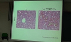
방사광 X-선 현미경은 임상실험에 유용한 도구로 높은 배율과 고 해상도로 동물 장기조직 시료의 세부 구 조를 관찰할 수 있다. X-선 영상은 위상 대조도 메커니즘으로 설명할 수 있다. 우리...
다국어 입력
あ
ぁ
か
が
さ
ざ
た
だ
な
は
ば
ぱ
ま
や
ゃ
ら
わ
ゎ
ん
い
ぃ
き
ぎ
し
じ
ち
ぢ
に
ひ
び
ぴ
み
り
う
ぅ
く
ぐ
す
ず
つ
づ
っ
ぬ
ふ
ぶ
ぷ
む
ゆ
ゅ
る
え
ぇ
け
げ
せ
ぜ
て
で
ね
へ
べ
ぺ
め
れ
お
ぉ
こ
ご
そ
ぞ
と
ど
の
ほ
ぼ
ぽ
も
よ
ょ
ろ
を
ア
ァ
カ
サ
ザ
タ
ダ
ナ
ハ
バ
パ
マ
ヤ
ャ
ラ
ワ
ヮ
ン
イ
ィ
キ
ギ
シ
ジ
チ
ヂ
ニ
ヒ
ビ
ピ
ミ
リ
ウ
ゥ
ク
グ
ス
ズ
ツ
ヅ
ッ
ヌ
フ
ブ
プ
ム
ユ
ュ
ル
エ
ェ
ケ
ゲ
セ
ゼ
テ
デ
ヘ
ベ
ペ
メ
レ
オ
ォ
コ
ゴ
ソ
ゾ
ト
ド
ノ
ホ
ボ
ポ
モ
ヨ
ョ
ロ
ヲ
―
http://chineseinput.net/에서 pinyin(병음)방식으로 중국어를 변환할 수 있습니다.
변환된 중국어를 복사하여 사용하시면 됩니다.
예시)
- 中文 을 입력하시려면 zhongwen을 입력하시고 space를누르시면됩니다.
- 北京 을 입력하시려면 beijing을 입력하시고 space를 누르시면 됩니다.
А
Б
В
Г
Д
Е
Ё
Ж
З
И
Й
К
Л
М
Н
О
П
Р
С
Т
У
Ф
Х
Ц
Ч
Ш
Щ
Ъ
Ы
Ь
Э
Ю
Я
а
б
в
г
д
е
ё
ж
з
и
й
к
л
м
н
о
п
р
с
т
у
ф
х
ц
ч
ш
щ
ъ
ы
ь
э
ю
я
′
″
℃
Å
¢
£
¥
¤
℉
‰
$
%
F
₩
㎕
㎖
㎗
ℓ
㎘
㏄
㎣
㎤
㎥
㎦
㎙
㎚
㎛
㎜
㎝
㎞
㎟
㎠
㎡
㎢
㏊
㎍
㎎
㎏
㏏
㎈
㎉
㏈
㎧
㎨
㎰
㎱
㎲
㎳
㎴
㎵
㎶
㎷
㎸
㎹
㎀
㎁
㎂
㎃
㎄
㎺
㎻
㎽
㎾
㎿
㎐
㎑
㎒
㎓
㎔
Ω
㏀
㏁
㎊
㎋
㎌
㏖
㏅
㎭
㎮
㎯
㏛
㎩
㎪
㎫
㎬
㏝
㏐
㏓
㏃
㏉
㏜
㏆

RISS 인기검색어
https://www.riss.kr/link?id=A100174233
- 저자
- 발행기관
- 학술지명
- 권호사항
-
발행연도
2008
-
작성언어
Korean
-
주제어
Synchrotron radiation ; Mouse tail ; Nerve ; Lung ; X-ray
-
KDC
512
-
자료형태
학술저널
-
수록면
23-30(8쪽)
-
KCI 피인용횟수
0
- 제공처
-
0
상세조회 -
0
다운로드
부가정보
국문 초록 (Abstract)

방사광 X-선 현미경은 임상실험에 유용한 도구로 높은 배율과 고 해상도로 동물 장기조직 시료의 세부 구 조를 관찰할 수 있다. X-선 영상은 위상 대조도 메커니즘으로 설명할 수 있다. 우리는 쥐의 꼬리, 신경 및 허파 의 in-vivo 및 in-vitro위상 대조도 영상을 8 KeV mono 빔으로부터 10배 현미경대물렌즈와 CCD 카메라를 이용하여 얻었다. 기존의 흡수 X-선 영상 보다 SR 영상이 세밀한 구조의 높은 분해능 영상을 볼 수 있었다. SR 영상은 생물학, 재료 및 임상 연구에 무한한 가능성을 가지고 있다.
방사광 X-선 현미경은 임상실험에 유용한 도구로 높은 배율과 고 해상도로 동물 장기조직 시료의 세부 구 조를 관찰할 수 있다. X-선 영상은 위상 대조도 메커니즘으로 설명할 수 있다. 우리는 쥐의 꼬리, 신경 및 허파 의 in-vivo 및 in-vitro위상 대조도 영상을 8 KeV mono 빔으로부터 10배 현미경대물렌즈와 CCD 카메라를 이용하여 얻었다. 기존의 흡수 X-선 영상 보다 SR 영상이 세밀한 구조의 높은 분해능 영상을 볼 수 있었다. SR 영상은 생물학, 재료 및 임상 연구에 무한한 가능성을 가지고 있다.
다국어 초록 (Multilingual Abstract)
X-ray microscopy with synchrotron radiation(SR) might be a useful tool for novel x-ray imaging in the clinical and laboratory settings. Microscopically, it enables us to observe detailed structure of animal organs samples with a great magnification power and an excellent resolution. The phase contrast mechanisms in image by X-ray are described. The phase-contrast X-ray imaging with SR from in-vivo and in-vitro mouse tail, rat nerve and rat lung were obtained with an 8 KeV monochromatic beam. The visual image was magnified using 10x microscope objective lens and captured using an digital CCD camera. The results showed more structural details and high resolution images with SR imaging system than conventional X-ray radiography system. The SR imaging system may have a potential for imaging in biological researches, material applications and clinical radiography.
X-ray microscopy with synchrotron radiation(SR) might be a useful tool for novel x-ray imaging in the clinical and laboratory settings. Microscopically, it enables us to observe detailed structure of animal organs samples with a great magnification po...
X-ray microscopy with synchrotron radiation(SR) might be a useful tool for novel x-ray imaging in the clinical and laboratory settings. Microscopically, it enables us to observe detailed structure of animal organs samples with a great magnification power and an excellent resolution. The phase contrast mechanisms in image by X-ray are described. The phase-contrast X-ray imaging with SR from in-vivo and in-vitro mouse tail, rat nerve and rat lung were obtained with an 8 KeV monochromatic beam. The visual image was magnified using 10x microscope objective lens and captured using an digital CCD camera. The results showed more structural details and high resolution images with SR imaging system than conventional X-ray radiography system. The SR imaging system may have a potential for imaging in biological researches, material applications and clinical radiography.
참고문헌 (Reference)
1 B. I. Kim, 71 : 443-447, 2008
2 S. Jheon, 69 : 656-659, 2006
3 이동녕, "방사광 과학입문" 청문각 2003
4 임근영, "동물 폐 표본에서의 해부학적 구조물과 병리적 병변: 미세 전산화단층촬영과 세절편 다중 검출기 전산화단층촬영에서의 발견능 비교" 대한영상의학회 54 (54): 385-391, 2006
5 Meuli R, "Synchrotron radiation in radiology: radiology techniques based on synchrotron sources" 14 : 1550-1560, 2004
6 Momose A, "Recent advances in X-ray phase imaging" 44 : 6355-6367, 2005
7 Kunii N, "Morphometric comparison of the human optic nerve fiber with various other human nerve fibers" 39 : 922-927, 1999
8 Longo R, "Morphological breast imaging: tomography and digital mammography with synchrotron radiation" 497 : 9-13, 2003
9 Fulvia Arfelli, "Mammography with synchrotron Radiation: Phase-detection techniques" 215 : 286-289, 2000
10 D. Chapman, "Diffration enhanced X-ray imaging" 42 : 2015-, 1997
11 Dance D. R, "Diagnostic radiology with X-ray, In The Physics of medical imaging" Institute of Physics Publishing 20-26, 1988
12 Weisheng Y, "Aerosol-induced lung injuries observed by synchrotron radiation X-ray phase-contrast imaging technique" 262 (262): 304-312, 2007
13 Chau W. K, "A morphometric study of optic axons regenerated in a sciatic nerve graft of adult rats" 16 : 105-116, 2000
동일학술지(권/호) 다른 논문
-
- 한국방사선학회
- 강보선
- 2008
-
- 한국방사선학회
- 임재동
- 2008
-
- 한국방사선학회
- 최규락
- 2008
-
저자장 자기공명영상시스템에서 선주사확산강조영상기법 개발
- 한국방사선학회
- 홍철표
- 2008
분석정보
인용정보 인용지수 설명보기
학술지 이력
| 연월일 | 이력구분 | 이력상세 | 등재구분 |
|---|---|---|---|
| 2026 | 평가예정 | 재인증평가 신청대상 (재인증) | |
| 2020-01-01 | 평가 | 등재학술지 유지 (재인증) |  |
| 2017-01-01 | 평가 | 등재학술지 선정 (계속평가) | |
| 2016-01-01 | 평가 | 등재후보학술지 유지 (계속평가) |  |
| 2015-01-01 | 평가 | 등재후보학술지 유지 (계속평가) | |
| 2013-01-01 | 평가 | 등재후보학술지 유지 (기타) | |
| 2012-01-01 | 평가 | 등재후보학술지 유지 (기타) | |
| 2011-02-28 | 학술지명변경 | 한글명 : 한국방사선학회 논문지 -> 한국방사선학회논문지 | |
| 2010-01-01 | 평가 | 등재후보학술지 선정 (신규평가) | |
| 2008-01-24 | 학회명변경 | 한글명 : 방사선학회 -> 한국방사선학회영문명 : The Society of Radiology -> The Korea Society of Radiology |
학술지 인용정보
| 기준연도 | WOS-KCI 통합IF(2년) | KCIF(2년) | KCIF(3년) |
|---|---|---|---|
| 2016 | 0.28 | 0.28 | 0.36 |
| KCIF(4년) | KCIF(5년) | 중심성지수(3년) | 즉시성지수 |
| 0.37 | 0.37 | 0.452 | 0.05 |


이 자료와 함께 이용한 RISS 자료
나만을 위한 추천자료


