 국립중앙도서관 "우편 복사 서비스"로 연결 됩니다.

 RISS
RISS
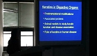
A 43-year-old man presented with dry cough and right chest pain. Chest X-ray showed normal and chest CT scan revealed thin-walled cysts of various sizes and nodules in both upper lobe. Abdominal CT scan revealed multiple low attenuated nodules in live...
다국어 입력
あ
ぁ
か
が
さ
ざ
た
だ
な
は
ば
ぱ
ま
や
ゃ
ら
わ
ゎ
ん
い
ぃ
き
ぎ
し
じ
ち
ぢ
に
ひ
び
ぴ
み
り
う
ぅ
く
ぐ
す
ず
つ
づ
っ
ぬ
ふ
ぶ
ぷ
む
ゆ
ゅ
る
え
ぇ
け
げ
せ
ぜ
て
で
ね
へ
べ
ぺ
め
れ
お
ぉ
こ
ご
そ
ぞ
と
ど
の
ほ
ぼ
ぽ
も
よ
ょ
ろ
を
ア
ァ
カ
サ
ザ
タ
ダ
ナ
ハ
バ
パ
マ
ヤ
ャ
ラ
ワ
ヮ
ン
イ
ィ
キ
ギ
シ
ジ
チ
ヂ
ニ
ヒ
ビ
ピ
ミ
リ
ウ
ゥ
ク
グ
ス
ズ
ツ
ヅ
ッ
ヌ
フ
ブ
プ
ム
ユ
ュ
ル
エ
ェ
ケ
ゲ
セ
ゼ
テ
デ
ヘ
ベ
ペ
メ
レ
オ
ォ
コ
ゴ
ソ
ゾ
ト
ド
ノ
ホ
ボ
ポ
モ
ヨ
ョ
ロ
ヲ
―
http://chineseinput.net/에서 pinyin(병음)방식으로 중국어를 변환할 수 있습니다.
변환된 중국어를 복사하여 사용하시면 됩니다.
예시)
- 中文 을 입력하시려면 zhongwen을 입력하시고 space를누르시면됩니다.
- 北京 을 입력하시려면 beijing을 입력하시고 space를 누르시면 됩니다.
А
Б
В
Г
Д
Е
Ё
Ж
З
И
Й
К
Л
М
Н
О
П
Р
С
Т
У
Ф
Х
Ц
Ч
Ш
Щ
Ъ
Ы
Ь
Э
Ю
Я
а
б
в
г
д
е
ё
ж
з
и
й
к
л
м
н
о
п
р
с
т
у
ф
х
ц
ч
ш
щ
ъ
ы
ь
э
ю
я
′
″
℃
Å
¢
£
¥
¤
℉
‰
$
%
F
₩
㎕
㎖
㎗
ℓ
㎘
㏄
㎣
㎤
㎥
㎦
㎙
㎚
㎛
㎜
㎝
㎞
㎟
㎠
㎡
㎢
㏊
㎍
㎎
㎏
㏏
㎈
㎉
㏈
㎧
㎨
㎰
㎱
㎲
㎳
㎴
㎵
㎶
㎷
㎸
㎹
㎀
㎁
㎂
㎃
㎄
㎺
㎻
㎽
㎾
㎿
㎐
㎑
㎒
㎓
㎔
Ω
㏀
㏁
㎊
㎋
㎌
㏖
㏅
㎭
㎮
㎯
㏛
㎩
㎪
㎫
㎬
㏝
㏐
㏓
㏃
㏉
㏜
㏆

RISS 인기검색어
부가정보
다국어 초록 (Multilingual Abstract)
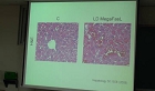
A 43-year-old man presented with dry cough and right chest pain. Chest X-ray showed normal and chest CT scan revealed thin-walled cysts of various sizes and nodules in both upper lobe. Abdominal CT scan revealed multiple low attenuated nodules in liver and multiple abodominal lymph nodes enlargement. Liver biopsy was performed. Light microscopic examination revealed proliferation and infiltration of Langerhans cell. Immunohistochemically, Langerhans cells showed strong cytoplasmic staining with S-100 protein and CD1a. He was diagnosed Langerhans' cell histiocytosis involving the lung and liver.
동일학술지(권/호) 다른 논문
-
요관경하 배석술 후 발생한 요관외벽에 함몰된 잔석 치료 경험
- 순천향의학연구소
- 이창호
- 2004
-
- 순천향의학연구소
- 한병규
- 2004
-
- 순천향의학연구소
- 유용덕
- 2004
-
무명정맥에서 상대정맥으로 이탈된 자가팽창형 stent의 경피적 제거
- 순천향의학연구소
- 구동억
- 2004
분석정보

이 자료와 함께 이용한 RISS 자료
나만을 위한 추천자료


