 국립중앙도서관 "우편 복사 서비스"로 연결 됩니다.

 ScienceON
목적 : 임상적으로 수지 사구종으로 의심되는 환자의 수술전 자기 공명 영상을 시행하여, 자기 공명 영상을 이용한 진단의 유용성과 변연 절제술 후의 임상적 결과를 알아보고자 하였다. 연...
다국어 입력
あ
ぁ
か
が
さ
ざ
た
だ
な
は
ば
ぱ
ま
や
ゃ
ら
わ
ゎ
ん
い
ぃ
き
ぎ
し
じ
ち
ぢ
に
ひ
び
ぴ
み
り
う
ぅ
く
ぐ
す
ず
つ
づ
っ
ぬ
ふ
ぶ
ぷ
む
ゆ
ゅ
る
え
ぇ
け
げ
せ
ぜ
て
で
ね
へ
べ
ぺ
め
れ
お
ぉ
こ
ご
そ
ぞ
と
ど
の
ほ
ぼ
ぽ
も
よ
ょ
ろ
を
ア
ァ
カ
サ
ザ
タ
ダ
ナ
ハ
バ
パ
マ
ヤ
ャ
ラ
ワ
ヮ
ン
イ
ィ
キ
ギ
シ
ジ
チ
ヂ
ニ
ヒ
ビ
ピ
ミ
リ
ウ
ゥ
ク
グ
ス
ズ
ツ
ヅ
ッ
ヌ
フ
ブ
プ
ム
ユ
ュ
ル
エ
ェ
ケ
ゲ
セ
ゼ
テ
デ
ヘ
ベ
ペ
メ
レ
オ
ォ
コ
ゴ
ソ
ゾ
ト
ド
ノ
ホ
ボ
ポ
モ
ヨ
ョ
ロ
ヲ
―
http://chineseinput.net/에서 pinyin(병음)방식으로 중국어를 변환할 수 있습니다.
변환된 중국어를 복사하여 사용하시면 됩니다.
예시)
- 中文 을 입력하시려면 zhongwen을 입력하시고 space를누르시면됩니다.
- 北京 을 입력하시려면 beijing을 입력하시고 space를 누르시면 됩니다.
А
Б
В
Г
Д
Е
Ё
Ж
З
И
Й
К
Л
М
Н
О
П
Р
С
Т
У
Ф
Х
Ц
Ч
Ш
Щ
Ъ
Ы
Ь
Э
Ю
Я
а
б
в
г
д
е
ё
ж
з
и
й
к
л
м
н
о
п
р
с
т
у
ф
х
ц
ч
ш
щ
ъ
ы
ь
э
ю
я
′
″
℃
Å
¢
£
¥
¤
℉
‰
$
%
F
₩
㎕
㎖
㎗
ℓ
㎘
㏄
㎣
㎤
㎥
㎦
㎙
㎚
㎛
㎜
㎝
㎞
㎟
㎠
㎡
㎢
㏊
㎍
㎎
㎏
㏏
㎈
㎉
㏈
㎧
㎨
㎰
㎱
㎲
㎳
㎴
㎵
㎶
㎷
㎸
㎹
㎀
㎁
㎂
㎃
㎄
㎺
㎻
㎽
㎾
㎿
㎐
㎑
㎒
㎓
㎔
Ω
㏀
㏁
㎊
㎋
㎌
㏖
㏅
㎭
㎮
㎯
㏛
㎩
㎪
㎫
㎬
㏝
㏐
㏓
㏃
㏉
㏜
㏆

RISS 인기검색어
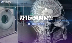
수지 조갑하 사구종의 진단 및 치료에서 자기 공명 영상의 임상적 의미 = Clinical Significance of MR Imaging for the Diagnosis and Treatment of Subungual Glomus Tumor in the Fingers
한글로보기https://www.riss.kr/link?id=A101528719
-
저자
김병석 ; 김우식 ; 한경진 ; 조재현 ; 이기범 ; 하헌교 ; 강신영 ; Kim, Byoung-Suck ; Kim, Woo-Sig ; Han, Kyoung-Jin ; Cho, Jae-Hyun ; Lee, Kyi-Beom ; Ha, Heon-Kyo ; Kang, Shin-Young
- 발행기관
- 학술지명
- 권호사항
-
발행연도
2001
-
작성언어
Korean
- 주제어
-
자료형태
학술저널
- 발행기관 URL
-
수록면
28-35(8쪽)
- 제공처
-
0
상세조회 -
0
다운로드
부가정보
국문 초록 (Abstract)
목적 : 임상적으로 수지 사구종으로 의심되는 환자의 수술전 자기 공명 영상을 시행하여, 자기 공명 영상을 이용한 진단의 유용성과 변연 절제술 후의 임상적 결과를 알아보고자 하였다. 연구대상 및 방법 : 수지 사구종을 의심하는 10례를 대상으로 수술전 문진, 이학적 검사, 단순 방사선 검사, 자기 공명 영상(9례)을 시행하였다. 수술적 치료는 변연 절제술을 시행하였으며, 수술후 병리 소견, 수술후 합병증 등을 비교 분석하였다. 결과 : 사구종으로 확진된 10례 중 호소하는 증세는 동통 10례, 압통 9례, 냉온에 대한 민감도 3례, 부종 1례였다. 자기 공명 영상 소견상 T1 강조 영상에서 저신호 강도 3례, 동신호 강도 5례, T2 강조 영상에서 고신호 강도 8례, 그리고 gadolinium 조영 증강된 영상에서는 8례 모두 조영 증가 소견을 보여 주었고, 발생 위치는 횡단면에서 정중부 6례, 외측부 5례, 조갑 외측 추벽 2례, 수지 두수 3례였고, 시상면에서 조갑상 5례, 조갑 기질 5례였다. 수술은 외측 접근법 1례, 조갑을 통한 접근법 9례를 시행하였으며, 모두 변연 절제술을 실시하였다. 수술후 전 예에서 임상 증세는 소실되었으며, 조갑 변형은 1례에서 발견되었으나, 재발은 없었다. 결론 : 수지의 사구종을 진단하는데 임상 증세는 아주 중요하나, 진단이 애매하거나 오랜 기간 동안 증세가 있어온 환자들에게서 비교적 비싼 비용을 지불하더라도 자기 공명 영상을 제한적으로 사용할 경우, 수술전 종물의 정확한 위치 확인 및 진단에 도움을 주는 방법의 하나로 생각한다.
다국어 초록 (Multilingual Abstract)
Purpose : Authors investigated the efficiency of preoperative MRI in suspicious glomus tumor and the clinical outcomes after marginal excision. Materials and Methods : In 10 cases of glomus tumors in the fingers, authors retrospectively analyzed the c...
Purpose : Authors investigated the efficiency of preoperative MRI in suspicious glomus tumor and the clinical outcomes after marginal excision. Materials and Methods : In 10 cases of glomus tumors in the fingers, authors retrospectively analyzed the clinical data, including previous trauma, treatment history, preoperative symptoms, physical examination, plain radiography, MRI (9 cases), pathological findings and postoperative complications. Results : The patients had pain in 10 cases, tenderness in 9 cases, cold sensitivity in 3 cases and edema in one case. MRI showed low signal (3 cases) or iso-signal (5) intensity on T1 weighted image, high signal intensity (8) on T2 weighted image, and all the lesions were enhanced in gadolinium enhancement images. The exact locations of glomus tumors were median in 6 cases, lateral in 5, lateral fold in 2 and pulp in 3 in transverse section and nail bed in 5 cases and nail matrix in 5 in sagittal section. Marginal excision was performed by lateral approach in one case and transungual in 9 cases. Histologically, all 10 cases were composed of solid sheets of round cells interrupted by thin-walled blood vessels. Most of clinical symptoms were disappeared in all cases after operation. Nail deformity was found in one case, which was originated from nail matrix, however, there was no recurrence. Summary : Clinical symptom was the most impotant factor in diagnosis of glomus tumor in the fingers. However, preoperative MRIs were helpful in patients, who had obscure pain or prolonged clinical symptoms with suspicious glomus tumors. Preoperative MRI might be one of the most useful tools for establishing the exact diagnosis and detecting the location of glomus tumors, in spite of the relatively high expenses.
동일학술지(권/호) 다른 논문
-
- 대한근골격종양학회
- 이승구
- 2001
-
- 대한근골격종양학회
- 강호정
- 2001
-
- 대한근골격종양학회
- 김주오
- 2001
-
항암제 함유 골시멘트에서 유리되는 아드리아마이신의 세포 독성
- 대한근골격종양학회
- 장동욱
- 2001
분석정보
이 자료와 함께 이용한 RISS 자료
나만을 위한 추천자료


